The Story of the Electrocardiograph: Many Intense Memories
Introduction

Ryozo Noguchi, Former Special Advisor, Fukuda Denshi Co., Ltd.
In Japan, the electrocardiograph has been in existence for almost 82 years, and electrocardiography is now a basic, indispensable test for the diagnosis of cardiac diseases.
Fukuda Denshi was also celebrating its 82nd anniversary since it was founded by the late Takashi Fukuda in 1939. Our path has focused exclusively on electrocardiographs, which medical specialists have helped us to develop. We have nothing but gratitude for their unfailing support and patronage.
In the following passages I describe some scenes in the history of electrocardiographs. I am honored to do so, as the history of electrocardiographs is ipso facto the history of our company, a “child of the electrocardiograph.” I have included episodes about the initial struggles faced by our doctors, and hope I can convey some measure of the intensity and energy which defined our early days.
Electrocardiographs are amazing
With measuring instruments, reproducibility is a major indicator of data reliability. With electrocardiographs, all that is required is to attach the electrodes properly, and the same waveform is reproduced time after time. While ultrasound diagnostic devices are also excellent testing devices, they are extremely operator-dependent, as the photographic data changes according to how the probe is set against the skin. By contrast, electrocardiographs have a major advantage in that it is easy to obtain the same waveform, regardless of who is operating the device. This is due to electrocardiographs having been developed on the basis of solid electrophysiological theory. The reliability and objectivity of the data were imperative for the establishment of present-day electrocardiographic theory.
The electrocardiograph is outstanding in its convenience, non-invasiveness, and capability of producing objective information. In the world of medical devices, it was a once-in-a-century masterpiece and a phenomenal invention of the 20th century, on a par with the x-ray machine.
Development of the photographic electrocardiograph: The advent of clinical application
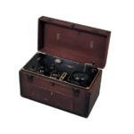
Photograph 1: The oldest domestically manufactured electrocardiograph in existence
The first prototype of electrocardiograph was produced in 1934. The initial electrocardiographs (photograph 1) were erratic, and even the staff at university hospitals were reduced to praying to the household altar to grant them a successful day of electrocardiography. From this pre-war infancy, the electrocardiograph began to blossom in the post-war period from around 1947-48. The electrocardiographs at the time were single-element, direct-current photographic instruments which recorded onto photosensitive oscillographic paper which was then developed. They were extremely simple devices: battery-operated, with 3-vacuum tube amplifier, moving iron type galvanometer, and spring motor. Subsequently, AC devices were developed using differential amplifiers to remove AC noise, and the momentum for clinical application finally began to grow. However, in the initial stages, the device had to be used in a bird cage-like, shielded room to combat AC noise. In addition, the instruments in those days broke down frequently, and there were many instances in which doctors themselves had to repair the galvanometer mirror and patient cables. There were also many times when the oscillographic paper only show a totally blackened image because of light which inadvertently found its way in during photography. It was only after this era of hand-made photography that the heated stylus, direct writing electrocardiograph was developed, paving the way for full-scale application to the clinical setting. In 1952, the Electrocardiograph Society a collaborative effort by medical scientists, engineers, and manufacturers, was established for the purpose of improving the devices. The Council played a major role in subsequent advancements in technology.
Development of the heated stylus direct writing electrocardiograph: On the way to a routine part of clinical testing
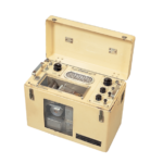
Photograph 2: The first domestically manufactured AC heated stylus direct writing electrocardiograph RS-1, produced in 1951
In 1951, Takashi Fukuda produced a prototype (Photograph 2) for the nation’s first heated stylus direct writing electrocardiograph (RS-1), modeled after an American direct writing electrocardiograph. Direct writing electrocardiographs were highly convenient due to their ability to record electrocardiograms on site. However, they were plagued by poor frequency response resulting in markedly inferior fidelity in high frequency ranges compared to photography type devices. The QRS complex in the electrocardiogram might be too low, or be missing nodes, or have a shallow Q wave.
From around 1957, the frequency response of heated stylus types began to show gradual improvement, and the use of electrocardiographs began to spread. Even so, the debate between photography and heated stylus raged on in academic circles in the early days, even serving as the theme for doctoral dissertations.
By about 1960 the frequency response of heated stylus types had improved to 50-60 c/s, with better waveform fidelity. There were also other issues, such as the galvanometer’s return to zero problem which had subtle effects on the ST-T waveform, the amplitude characteristics, and issues with domestically produced recording paper, but all of the manufacturers worked feverishly to solve these problems under the guidance of medical experts and scientists, collectively succeeding in improving the level of accuracy and performance of the heated stylus electrocardiograph. This led to the full-fledged incorporation of these devices into hospitals and clinics as a routine part of clinical examinations.
The natural enemy of electrocardiographs
The problem with AC powered devices was stabilizing the AC power source and removing AC noise due to interference. AC noise, in particular, is the natural enemy of electrocardiographs, and the biggest challenge was to extract accurate ECG waveforms while excluding noise. This problem was solved with the development of differential amplifiers, which cancel out noise and high performance hum filters.
I am sure many doctors remember struggling with hum, muscle noise, and baseline drift when recording ECGs. They had to work desperately just to perform the test, using shielding sheets, moving the bed, soothing the anxious patient, and so on.
In one case of stubborn noise that refused to go away, we found that the stylus was crooning the Japanese version of a country-and-western song. It turned out that the radio waves from a nearby broadcasting station found their way in via the cables. This is just one example which illustrates how the history of electrocardiographs was a continuous battle with its natural enemy: noise.
Classes on recording and reading electrocardiograms
From about 1955, Japan Medical Association began to give classes on how to record and read electrocardiograms. After all, this was a brand new testing methodology and doctors were very enthusiastic on mastering how to read electrocardiograms as quickly as they could. Instructors came from universities and the classes would start at 8 p.m. or so, after the doctors’ consultation work was finished, and frequently go on till late at night. Even so, the classes were attended by large numbers of doctors every time, and the lecturers gave their full support despite the strain on their workloads in order to spread the use of the electrocardiograph.
Doctors in Japan Medical Association also began to use the electrocardiograph more and more frequently in community and school health checkups. At the time, you could almost feel the passion radiating from the doctors in their commitment to rebuilding the nation.
Smaller, more lightweight devices
Transistors were incorporated to improve stability, durability, and other performance parameters, as well as to make electrocardiographs smaller, lighter, and more energy efficient. While problem-laden in the initial stages, such as in variable quality, temperature-induced property changes, and transistor wire breakage, the quality and performance of semiconductors underwent a quantum improvement with the domestic production of the field-effect transistor. This resulted in the development of the true IC electrocardiograph in 1966. With its transformers replaced by small, powerful, stabilized power sources, the electrocardiograph underwent a dramatic reduction in size and weight, while offering even better performance. The next step in its evolution would be microcomputer-based electrocardiograph.
Development of the microcomputer-based electrocardiograph
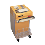
Photograph 3: Japan’s first microcomputer-based electrocardiograph FCP-30, developed in 1978
In the 1960s, research and experimentation on the automatic analysis of electrocardiograms were carried out in the United States and, with the evolution of the minicomputer, put into practical use. In Japan, too, the long years of study by doctors and engineers bore fruit in 1971, when these systems were employed in health checkups. At the time, analysis precision was still an issue with minicomputer systems, and the data had to be re-read by experts.
Advances in computer technology gave birth to the microcomputer, which offered performance on a par with minicomputers at a fraction of the size, and in 1978 IBM launched a microcomputer-based ECG acquisition and analysis system. In December of that same year, Fukuda Denshi launched the FCP-30, Japan’s first microcomputer-based electrocardiograph (Photograph 3). It was large, weighing 75 kg, and was a transitional product consisting of merely attaching a microcomputer to an existing electrocardiograph and mounting a printer capable of printing out the analysis results. Even so, it could be described as revolutionary.
Later, when minicomputer software was ported to microcomputers under the guidance of doctors, analysis precision improved dramatically. The units themselves also became smaller and digitized, as microcomputers were incorporated into conventional electrocardiographs. With advances in private sector thermal printing technology, electrocardiographs were equipped with thermal mechanisms, enabling simultaneous recording of ECG waveforms, patient information, and analysis results, printed out as a report onto A4 sized paper.
In recent developments, the devices have been equipped with Liquid Crystal Displays for both waveforms and operation panels, further improving user friendliness. They are now more ubiquitous than the conventional electrocardiographs and are in wide use in universities and major hospitals.
Development of automatic ECG transmission and analysis systems
In the early days, the technologically inferior software and faulty analysis programs of microcomputer-based electrocardiographs resulted in a significant amount of erroneous analysis results. This problematic analysis precision resulted in the need for more precise analysis and for overreading (checking of waveforms) by specialists. Fukuda Denshi swiftly embarked on the development of a digital ECG transmission and analysis system with overreading function using Fujitsu minicomputer and ordinary telephone lines.
In 1981, the first model of the system (FCP-1000) was delivered to the Osaka Medical Cooperative Association on recommendation by the Osaka Medical Association. The system featured improved waveform quality due to digitized transmission, better analysis reliability than microcomputer, and added overreading function. The overreading was performed at Kansai Medical University and Kinki University Hospital. Approximately 300 terminals were installed and operated on a 24-hour basis to achieve remarkable results.
Medical associations, universities, and key hospitals worked together to set up the Osaka Medical Association Committee on Automatic ECG Analysis, and the Guidebook on Automatic ECG Analysis was created. It was an outstanding guidebook providing user-friendly commentary of analysis findings, and was actively utilized on a nationwide basis. It was one of the first models for collaboration between doctors and universities.
By 1985, an offline system for automatic analysis of ECG and PCG of school-age children was added and became operative, enhancing the efficiency of the system. Subsequently, this system was delivered to regional medical associations including those in Kagoshima and Okayama Prefectures, as well as to universities and key hospitals such as Kanazawa Medical University, Shimane Medical University, and Kurashiki Central Hospital, where it produced high levels of performance.
In the present day, this system has been superseded by the microcomputer-based electrocardiograph. Even so, the technologies and know-how will surely be useful in internet-based ECG data network systems.
Precision and reliability of automatic analysis
Photograph 4: Electrocardiograph with analysis function, FCP-3610
Automatic analysis uses methods similar to those a doctor uses to read ECGs. The ECG waveform input is digitized (A/D conversion), and noise and drift are corrected (pre-processing). The waveforms are then measured and analyzed.
The precision and reliability of analysis are dependent on noise removal, waveform recognition, and measurement precision. Today, analysis precision has improved significantly, at about 95% in health checkups and 80% in clinical settings. Diagnosis of normal range can be described as definite, and reliability is believed to be equivalent to that of a specialist with several years’ experience.
The CSE database for waveform measurement and diagnosis is widely accepted as the world standard, and is used for comparing the measurement and diagnostic precision of products by companies around the world. Fukuda Denshi is the only Japanese company participating in this study, and our analysis precision has been demonstrated to be on a par with that of IBM, Telemed, and HP.
In automatic analysis, the rationale for analysis, that is, the reason behind the analysis, is important. At Fukuda Denshi, we recently developed and incorporated into our microcomputer-based electrocardiographs, a program to provide analysis guidance, to identify the rationale for analysis on patient-specific findings. We hope this will further promote understanding of automatic analysis.
Conclusion
The development of electrocardiographs was largely dependent on the advances in private sector electronic devices, with particularly spectacular changes occurring with leaps in digital technology associated with the advent of the computer. Even so, the advances would not have been possible without the specific know-how on technology, development, and production of electrodes, galvanometers, and analysis programs. Going forward, these kinds of technologies will continue to be added as the devices continue their evolution. Furthermore, electrocardiographs came this far because engineers worked under the guidance of doctors. Medical engineering will never exist in the absence of clinical practice, and we will continue to look to doctors for guidance. Our aging population also necessitates more electrocardiographic management of day to day lives. To meet this need, it is likely that super-small, single-chip electrocardiographs will be developed. Electronic patient records applying ECG filing technologies will likewise become more widespread. In the context of these developments, Fukuda Denshi is committed to contributing to medical science through medical engineering equipment, capitalizing on our specialized manufacturing technology. We continue to look to doctors for feedback and support. Hitherto I have limited my discussions to a concise overview, omitting many details for the sake of brevity. If this has compromised lucidity in any way I humbly ask for the reader’s understanding. Thank you for your attention.